What Is Stereotactic Biopsy
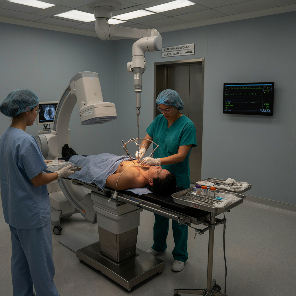
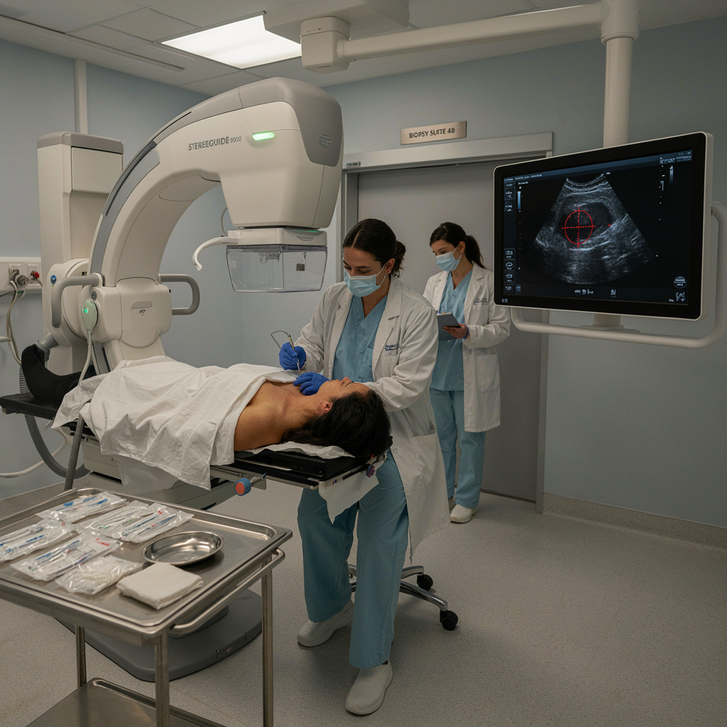
Stereotactic biopsy is an advanced image-guided procedure used to obtain a small tissue sample from a brain or other deep-seated lesion. Dr. Aneel Kumar P uses detailed CT or MRI scans and precise computer-assisted navigation to carefully guide a biopsy needle to the targeted area. This minimally invasive technique helps obtain accurate tissue samples for diagnosis while minimizing damage to surrounding healthy brain tissue. It is commonly used to identify tumors, infections, or other abnormalities and helps doctors determine the most appropriate treatment plan.
- Precise image-guided targeting
- Minimally invasive tissue sampling
- Accurate pathological diagnosis
- Reduced surgical tissue disruption
Advanced Neuro Oncology Care
Comprehensive, precision-focused brain tumor treatments combining advanced technology, surgical expertise, and personalized care for safer outcomes.
How the Procedure Works
Dr. Aneel Kumar P uses advanced imaging to help accurately identify the target and safely obtain tissue for laboratory examination.
Why Choose Stereotactic Biopsy
Dr. Aneel Kumar P provides advanced, image-guided stereotactic biopsy focused on obtaining accurate tissue samples from deep or difficult-to-reach lesions. His approach emphasizes precise targeting, minimal tissue disruption, diagnostic accuracy, and effective treatment planning.
Frequently Asked Questions
The procedure duration varies depending on the lesion location, imaging requirements, and individual patient circumstances.
It is generally considered a safe minimally invasive procedure, although risks depend on the lesion location and individual health factors.
Accuracy is generally high because imaging guidance helps the surgeon target the specific area requiring tissue sampling.
Preliminary findings may sometimes be available quickly, while complete pathological analysis can take additional time depending on the tests required.
Patients are monitored after the procedure and receive individualized instructions regarding observation, wound care, activity, and follow-up.